Phone : +91 40-2403 8699 / 8799
Mobile : +91 95151 34669
Email : urmiladiagnostics369@gmail.com

Phone : +91 40-2403 8699 / 8799
Mobile : +91 95151 34669
Email : urmiladiagnostics369@gmail.com
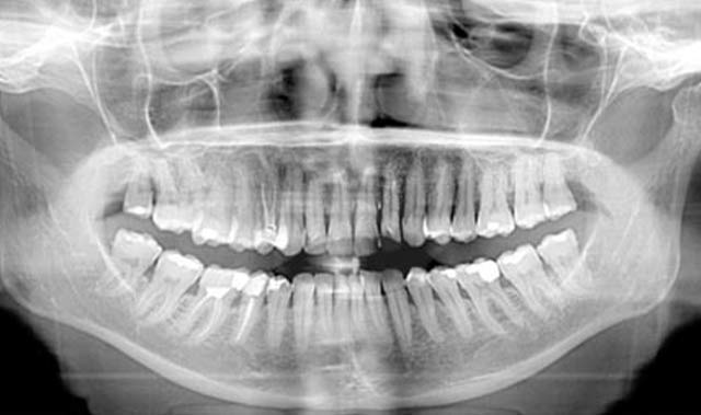
This x-ray scan illustrates the maxilla and mandible, all the teeth including the "wisdom teeth," the frontal, nasal and maxillary sinus air spaces and the temporomandibular joint complex or "TMJ".
An Orthopantomogram (OPG), also known as an "orthopantogram" or "panorex", is a panoramic scanning dental X-ray of the upper and lower jaw. It shows a two-dimensional view of a half-circle from ear to ear.
Equipment Dental panoramic radiography equipment consists of a horizontal rotating arm which holds an X-ray source and a moving film mechanism (carrying a film) arranged at opposed extremities. The patient's skull sits between the X-ray generator and the film. The X-ray source is collimated toward the film, to give a beam shaped as a vertical blade having a width of 4-7mm when arriving on the film, after crossing the patient's skull. Also the height of that beam covers the mandibles and the maxilla regions. The arm moves and its movement may be described as a rotation around an instant centre which shifts on a dedicated trajectory.
The manufacturers propose different solutions for moving the arm, trying to maintain constant distance between the teeth to the film and generator. Also those moving solutions try to project the teeth arch as orthogonally as possible. It is impossible to select an ideal movement as the anatomy varies very much from person to person. Finally a compromise is selected by each manufacturer and results in magnification factors which vary strongly along the film (15%-30%). The patient positioning is very critical in regard to both sharpness and distortions.
The result is an image showing sharply the section along the mandible arch and blurred the rest. For example, the more radio-opaque anatomical region, the cervical vertebras (neck), shows as a wide and blurred vertical pillar overlapping the front teeth. The path where the anatomical elements are recorded sharply is called "focal path".
Dental X-rays radiology moves from film technology (involving a chemical developing process) to Digital X-ray which is based on electronic sensors and computers. One of the principal advantages compared to film based systems is the much greater exposure latitude. This means much less repeats, which also reduces patient exposure to radiation. Lost xrays can also be reprinted. Other significant advantages include instantly viewable images, ability to enhance images, ability to email images to practitioners and clients, easy and reliable document handling, reduced X-ray exposure, no darkroom is required, no chemicals are used.
One particular type of digital system uses a Photostimulable Phosphor Plate (aka PSP - Phosphor Plate) in place of the film. After X-ray exposure the plate (sheet) is placed in a special scanner where the latent formed image is retrieved point by point and digitized, using a laser light scanning. The digitized images are stored and displayed on the computer screen. This method is half way between old film based technology and the current direct digital imaging technology. It is similar to the film process because it involves the same image support handling and differs because the chemical development process is replaced by the scanning process. This is not much faster that film processing and the resolution and sensitivity performances are contested. However it has the clear advantage to be able to fit with any pre-existing equipment without any modification because it replaces just the existing film. Also some times the term "Digital X-rays" is used to designate the scanned film documents which further are handled by computers. In current state-of-the-art digital systems, the image quality is vastly superior to conventional film-based systems.
OPGs are used by Dentists to provide information on :
The most common use is to determine the status of wisdom teeth.